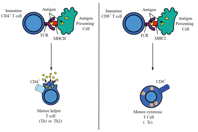
The first step is an activation of the immature T-Cells to mature cells (Transduction) by contact with Antigen Presenting Cells and co-stimulation molecules. The activation is shown when Interleukins are produced and a cell volume shift is then observed. Experts can now genetically modify the immune cells by transferring relevant gene (Transfection).
Your challenges
- How can I track volume shift of cells during activation process?
- Is your multi-parametric flow cytometry method robust enough to assess accurate gene expression & phenotype & at the same time recording data in compliance with 21 CFR part 11?
- Are you concerned by biosafety or sample contamination?
-
Do the preparation purety & cell recovery rates impact your process?
Understanding the Role and Activation Mechanism of CD4+ T Cells in Immune Response
Activation of CD4+ T cells occurs through the simultaneous engagement of the T-cell receptor (TCR) and a co-stimulatory molecule (like CD28, or ICOS) on the T cell by the major histocompatibility complex (MHCII) peptide & co-stimulatory molecules on the antigen-presenting cell (APC). Both are required for production of an effective immune response; in the absence of co-stimulation, T cell receptor signalling alone results in anergy. Optimal CD8+ T cell response relies on CD4+ signalling.[1] CD4+ cells are useful in the initial antigenic activation of naive CD8 T cells, and sustaining memory CD8+ T cells in the aftermath of an acute infection. Therefore, activation of CD4+ T cells can be beneficial to the action of CD8+ T cells. [2][3][4]
The first signal is provided by binding of the T cell receptor to its cognate peptide presented on MHCII on an APC. MHCII is restricted to so-called professional antigen-presenting cells (APC), like dendritic cells, B cells, and macrophages, to name a few.
 Antigen presentation stimulates T cells to activate "cytotoxic" CD8+ cells or "helper" CD4+ cells. Cytotoxic cells directly attack other cells carrying certain foreign or abnormal molecules on their surfaces. Helper T cells, or Th cells, coordinate immune responses by communicating with other cells. In most cases, T cells only recognize an antigen if it is carried on the surface of a cell by one of the body’s own MHC, or major histocompatibility complex, molecules.
Definitions
Signal transduction
Signal transduction is by definition the conversion of a signal from one form to another.
For lymphocytes, signal transduction begins at the plasma membrane and is initiated by the binding of antigen (foreign substances or microorganisms that the host recognizes as “nonself”) to the T cell receptor (TCR) or the B cell receptor (BCR).
As a result of this binding the activation of several signalling cascades occurs, resulting in the propagation and expansion of the initial signal. For lymphocytes, ultimately the response to extracellular signals is the induction of a new gene transcription patter
Transfection
Transfection is the process of deliberately introducing naked or purified nucleic acids into eukaryotic cells
The word transfection is a portmanteau of trans- and infection.
Genetic material (such as supercoiled plasmid DNA or siRNA constructs), or even proteins such as antibodies, may be transfected.
Transfection of animal cells typically involves opening transient pores or "holes" in the cell membrane to allow the uptake of material. Transfection can be carried out using calcium phosphate (i.e. tricalcium phosphate), by electroporation, by cell squeezing or by mixing a cationic lipid with the material to produce liposomes that fuse with the cell membrane and deposit their cargo inside.
Sources and references
Picture source By user:Sjef - Own work (Original text: self made,
http://commons.wikimedia.org/wiki/Image:Antigen_presentation.jpg),
CC BY-SA 3.0,
https://commons.wikimedia.org/w/index.php?curid=4470656.
[1] Williams, M. A., & Bevan, M. J. (2007). Effector and memory CTL differentiation. Annual review of immunology, 25, 171–192.
https://doi.org/10.1146/annurev.immunol.25.022106.141548
[2] Janssen, E., Lemmens, E., Wolfe, T. et al. (2003). CD4+ T cells are required for secondary expansion and memory in CD8+ T lymphocytes. Nature 421, 852–856.
https://doi.org/10.1038/nature01441
[3] Shedlock, D. J., & Shen, H. (2003). Requirement for CD4 T cell help in generating functional CD8 T cell memory. Science (New York, N.Y.), 300(5617), 337–339.
https://doi.org/10.1126/science.1082305
[4] Sun, J. C., Williams, M. A., & Bevan, M. J. (2004). CD4+ T cells are required for the maintenance, not programming, of memory CD8+ T cells after acute infection. Nature immunology, 5(9), 927–933.
https://doi.org/10.1038/ni1105
[5] Brundage K.M. (2005) Signal Transduction During Lymphocyte Activation. In: Vohr HW. (eds) Encyclopedic Reference of Immunotoxicology. Springer, Berlin, Heidelberg.
https://doi.org/10.1007/3-540-27806-0_1350
Harris, K. (2019, June 23). Signal Transduction. Retrieved April 8, 2021, from
https://bio.libretexts.org/@go/page/23979